Pathology Slide Scanner

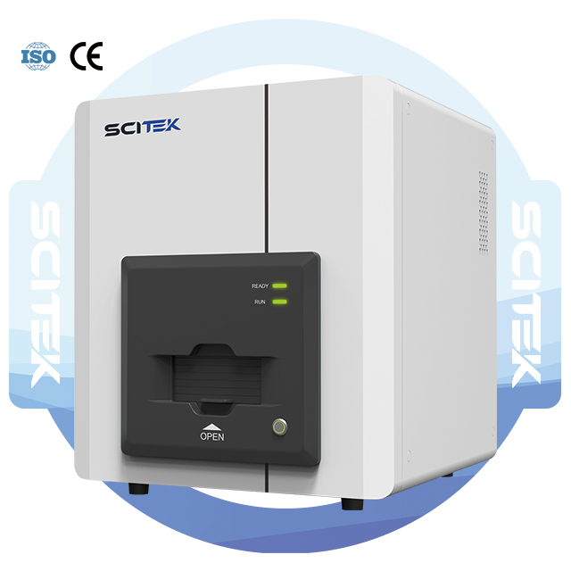
The pathology slide scanner is equipped with a magnetic design that ensures precise positioning and smooth loading/unloading.

  • Model : DPS-K 
  • Loading Capacity : 5 slides
  • Slide Size : 26×76mm
  • Objective Lens : Plan Apochromatic (APO) 20× objective, Numerical Aperture NA 0.75
  • Core Imaging Camera : 5-megapixel CMOS area-array camera, 2/3 inch

Share the product

分类: 标签:

Pathology Slide Scanner Advantage:

  • Segmented Power Control for Safe Operation
  • High and low voltage circuits are independently controlled during startup to prevent “surge” damage to sensitive electronic modules and ensure operator safety.


  • Magnetic Slide Holder
  • Magnetic design ensures precise positioning and smooth loading/unloading, preventing slide damage or slippage.


  • High-Speed, High-Precision X/Y/Z Stage System
  • Imported magnetic levitation linear motor: Speed up to 3.2m/s, acceleration up to 8g.
  • Full closed-loop control system: X/Y-axis resolution 100nm, Z-axis resolution 50nm.


  • Advanced Scanning Software Features
  • Instant preview: View scanned images immediately for diagnosis.
  • Real-time positioning: Microscope-like navigation to locate and display scanned regions.
  • Dynamic parameter display: Shows real-time positional and tissue information.
  • Multi-tissue recognition: Intelligently segments and scans multiple tissue blocks on a single slide, merging them into a unified digital image.


  • Viewing Software Capabilities
  • Slide management: Automatically detects, sorts, searches, and categorizes slide files.
  • Color-coded navigation: Tracks viewing paths at different magnifications.
  • ROI annotation: Marks regions of interest with customizable shapes, colors, and line widths; exports annotations to Excel.
  • High-resolution capture: Supports 2300dpi screenshots (partial or full-screen) for publication; allows labeling and exporting.

Tissue Slide Dryer Specification:

Model DPS-K
Loading Capacity 5 slides
Slide Size 26mm×76mm
Objective Lens Plan Apochromatic (APO) 20× objective, Numerical Aperture NA0.75
Core Imaging Camera 5-megapixel CMOS area-array camera, 2/3 inch
Scan Magnification 20×/40× automatic switching
Scan Method Area-array scanning
Scan Area ≥24mm× 50mm
Scan Speed 20×: <30 seconds
Focusing Method Auto-focusing and manual fine-tuning
Specimen Identification Supports standard barcodes, QR codes, etc.
Tissue Recognition Automatically detects tissue shape and region; skips blank areas; intelligently segments and scans multiple tissue blocks on a single slide
Multi-layer Fusion Scan Stores individual layer images for cellular morphology observation; fuses clearest regions from each layer into a composite image
Scan Output 24-bit color, real-time preview
Data Storage Local and cloud storage
Shipping Size(W×D×H)(mm) 860×850×1450
Weight 153kg


标题

CONTACT INFO

No. 7000 Jingshi Road, Jinan City, Shandong Province, China

座机: +86-531-82893797

返回顶部